Description
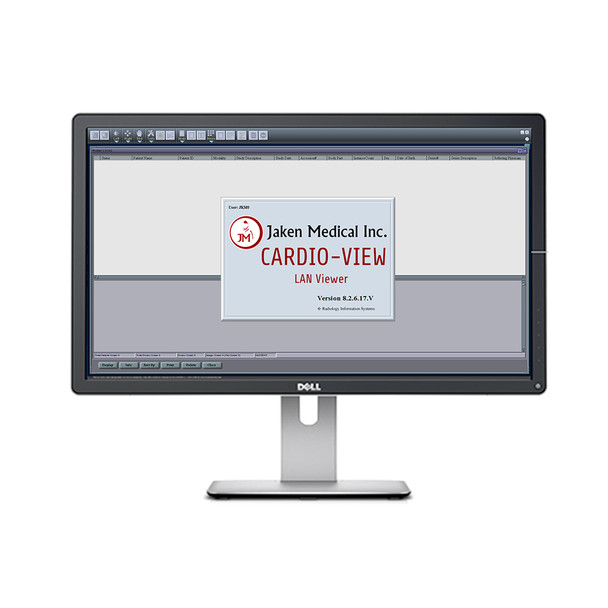
Jaken Cardio-View LAN DICOM Viewing Software (Single User)
Download 30-Day Trial Software [Direct Download]
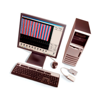
Jaken Cardio-View DICOM Viewing, Reporting & Archiving Solutions: For Cardiologists looking for a high-quality Viewer at an affordable, low cost. Download your Jaken Cardio-View 30-Day Trial Software Now!
Viewer Features:
- Reporting
- Study note creation
- Window/leveling
- Pan
- Zoom
- CD - Import
- CD - Burning (SWRSVSCGB)
- Windows print
- Shutter
- Magnifying glass
- Pseudo color
- Export image/series as DICOM, JPEG, BMP, AVI files
- Attach documents to study
- Attach non-dicom image to study (ie jpg)
- Receive studies from PACS via DICOM
- Query / Retrieve from PACS via DICOM
- Send images and attached annotations / documents to PACS via DICOM
- Rotate / Flip Orientation ICONS
- Information Text overlay (patient name / study info)
- Annotation graphics (line, curve, circle, text, arrow)
- Basic Measurement (distance, Area, Angle, Cobb angle, Density, angle of intersecting lines, Heart / Chest Ratio)
- EchoCardiology Measurement (Peak Pressure Gradient, Velocity Time Integration, LA volume Bi-Plane, Volume single plane, LV volume Modified Simpson's method Bi-Plane, Ejection Fraction, Deceleration Time, Diameter for Circular Area, Y0 Calibration)
- Multi-Frame viewing and comparison with playback tools
- Creates a Local "worklist" of all Studies and Patients for easy access and viewing
- Can compare studies side by side on same monitor
Viewer Echocardiography Tools:
- Peak Pressure Gradient:
- Velocity-Time Integration (VTI):
- Left Atrial Volume:
- Volume Measurement with Single-Plane Area-Length Method:
- Left Ventricular Volume:
- Ejection Fraction:
- Deceleration Time:
- Diameter for Circular Area:
- Y0 Calibration:
Echocardiography Ultrasound Image Regions:
Currently supported echocardiography measurement tools only apply to the following Ultrasound Image Regions.
- 2D regions, including 2D Tissue and 2D Color Flow regions.
- M-mode Tissue region and Doppler Velocity Waveform region.
The Doppler Velocity Waveform region allows the following measurement tools to be performed.
- Peak Pressure Gradient
- Velocity-Time Integration
- Deceleration Time
- Y0 Calibration
2D regions accept the following measurements.
- Volume Measurement: Single-Plane Area-Length method
- Volume Measurement: Biplane Area-Length method
- Volume Measurement: Modified Simpsonís Biplane method
- Ejection Fraction
2D regions and the M-mode Tissue region allow the following measurement.
- Diameter for Circular Area
Download 30-Day Trial Software [Direct Download]
- Viewing, Reporting & Archiving Solution
- High-quality viewer at an affordable price
- 30 Day Trial Software


